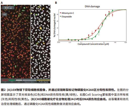
Foreword DNA or chromosome damage is often involved in research because DNA damage is closely related to the occurrence of many diseases, such as genetic diseases and tumors. Radiation radiation, environmental effects, or combined drugs can cause DNA damage. Previous studies have shown that the phosphorylation site of the labeled histone H2AX on serine 139 is a sensitive method for early prediction of DNA double-strand breaks. In this paper, the experimental method of detecting DNA damage by automatic cell imaging is described in detail. The cell sample is treated by small molecule compound and imaged by phosphorylated histone H2AX immunofluorescence staining. In addition, three cell lines, CHO, Hela and PC-12, were added to the experiment, and mitomycin and etoposide were added to detect the effects of the compounds. Phosphorylated H2AX immunofluorescence method for quantification of DNA damage Cell-level DNA damage can be visualized and quantified using commercial reagents and the ImageXpress ® Nano automated imaging and analysis system. The immunofluorescence method used a primary antibody against phosphorylated H2AX (EMD Millipore) and an AlexaFluor labeled secondary antibody (Life Technologies). Hoechst33342 was stained with nuclei, imaged by ImageXpress Nano, 10X objective, DAPI and FITC channels. - Seeding cells in a flat, transparent 384-well plate, 5,000-7,500 cells/well Advantage - After the cells are fixed, the nuclei are fluorescently stained by immunoassay and imaged. Cell Scoring Module for Real-Time and Efficient Quantification of DNA Damage DNA Damage CellReporterXpress TM automatic image acquisition and analysis software includes a preset Cell Scoring module, which can easily damage flag is set classified according to positive and negative DNA damage, and calculate the percentage of cells that produce DNA damage. Image acquisition and analysis are usually run together, and the image and analysis data can be seen simultaneously when the well plate is read. In addition, due to the large target surface characteristics of the detector, the cells in one field under the 10X objective were sufficiently quantified to obtain statistically significant concentration effect curve data (Fig. 2). Secondly, under this shooting condition, one field of view can capture >500 cells (maximal concentration of mitomycin C) and >2800 cells (mildicidal concentration of mitomycin C). references 1. Paull TT, Rogakou EP, Yamazaki V., Kirchgessner CU, Gellert M., BonnerW.M..2000. A critical role for histone H2AXin recruitment of repair factors to nuclearfoci after DNA damage. Curr Biol.
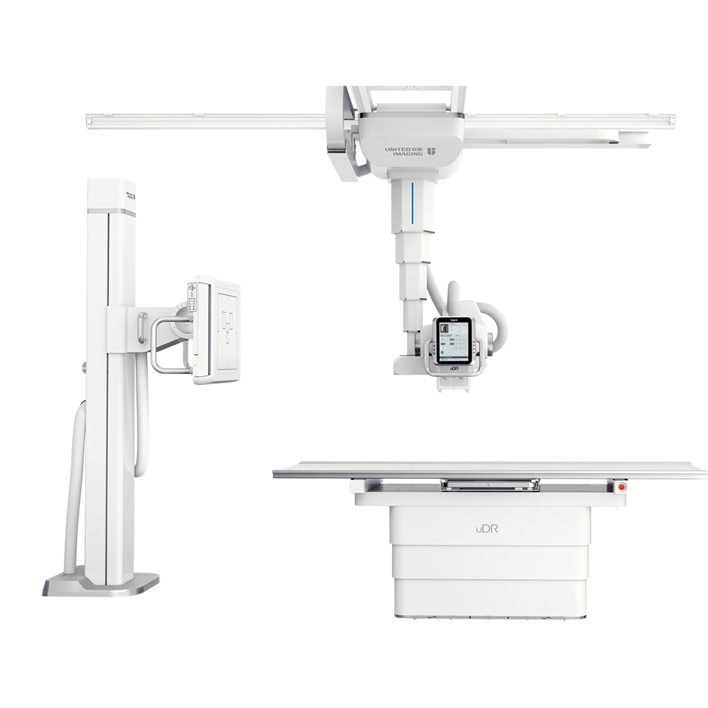
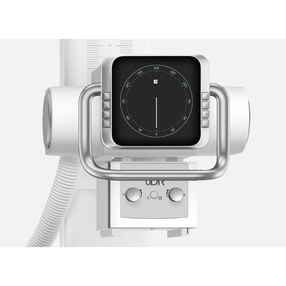
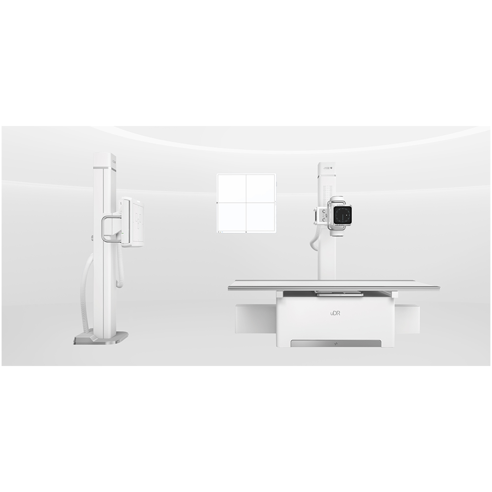
Digital Radiography made affordable, Created to meet the needs of community to hospitals and private radiology practices, it enables the price-sensitive customers to join the drive to go digital.The versatile floor mounted radiography system enables you to go from film to CR to DR. Flooe-mounted easy for installation and operation.
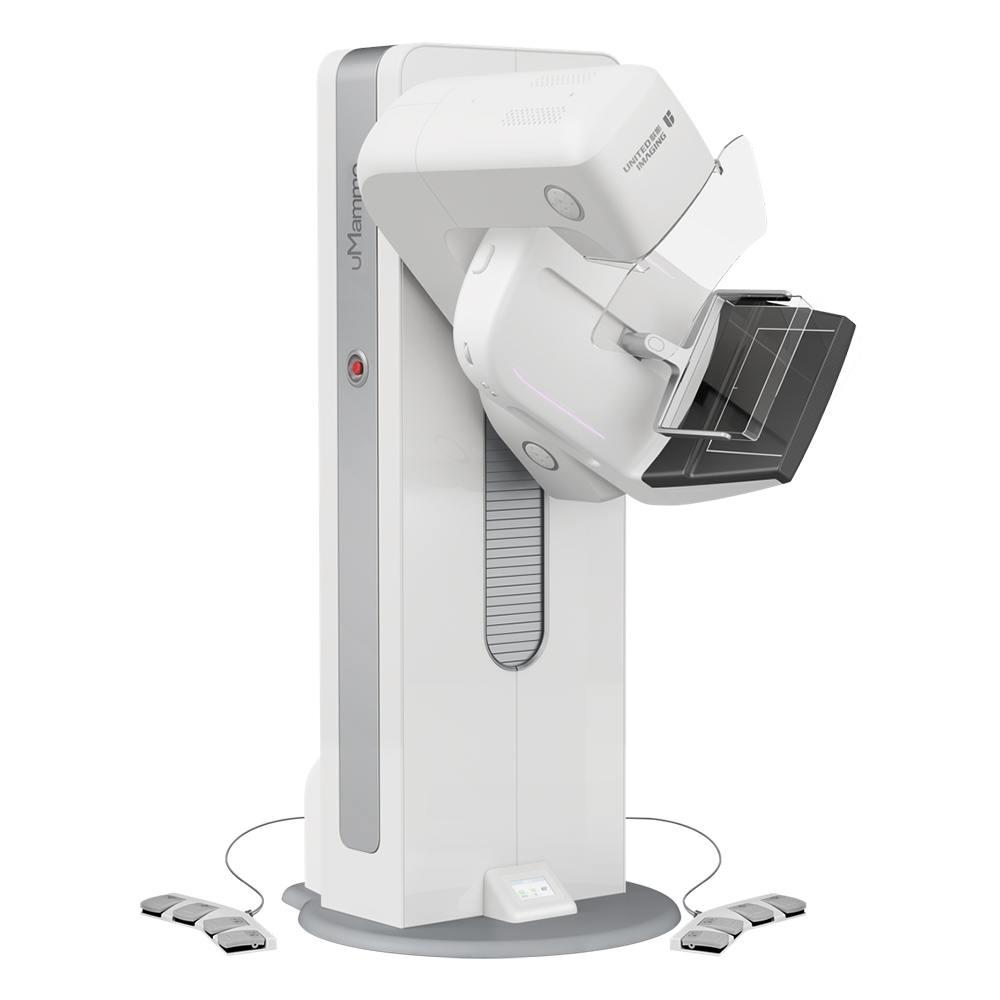
United-imaging according to the international advanced processing mode and standardlize design. the parts are made with precision CNC machine and moulding. Which adopt high degree of standardized, reasonable and compact structure. With reliable, durable and elegant appearance and advanced processing technology.
X-Ray Digital Machine,X-Ray Digital Machine In Medical,X-Rays Digital Medical Machine,Medical X-Ray Digital Imaging Machine Shanghai Rocatti Biotechnology Co.,Ltd , https://www.shljdmedical.com
- Place cells overnight at 37 ° C / 5% CO 2
- Add 1:2 series of gradient DNA toxic compounds to the wells and treat the cells for 18-24 hours
- Fix cells with 4% paraformaldehyde at room temperature
- Wash the fixative with PBS, then block and punch the cells with 1% BSA + 0.1% Triton X-100 and incubate for 30 minutes at room temperature.
- Add anti-H2AX primary antibody overnight at 4 °C
- Wash 3 times in 1XPBS
- Add fluorescent secondary antibody and 16μM Hoechst for 1-2 hours at room temperature
- Wash 3 times in 1XPBS
- Obtain images with the ImageXpressNano system 10X objective and run the CellScoring module for real-time quantification
- Accurate calculation of DNA double-strand break sites
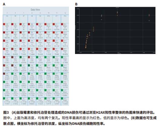
- On-the-fly analysis yields calculation results in real time
- Display positive holes by heat map 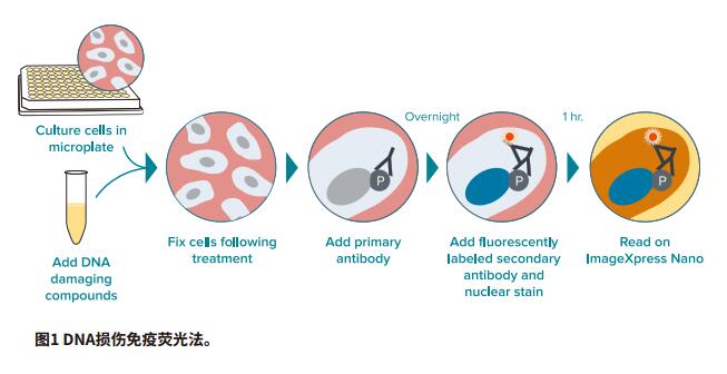

The United-imaging is floor mounted system comprises a radiographic table with integral floor guide rail and a wallstabd. Requiring little room preparation, it is easy to install.
